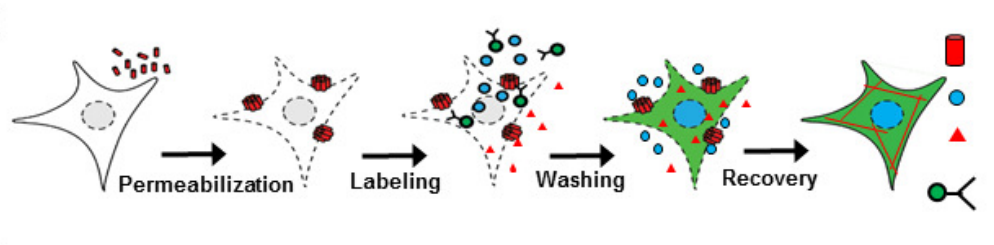
The technique of fluorescent microscopy is often based on the specific labeling of molecular inside the cell using fluorescent proteins or external fluorophores. However, most of antibodies and fluorophores are not able to across membrane of living cells. To overcome the impermeability of external probes, we develop a technique of intracellular labeling in living cells using Streptolysin O, a bacterial protein which can form pores on membrane. After transient permeablization, external antibodies and fluorophores can be delivered into cells, and then cells are able to repair their membrane. The whole process takes around 20 minutes and gives over 85% labeling efficiency. This technique opens the possibility to study living cells using various external fluorophores.
Related Publications:
- Kai Wen Teng, Yuji Ishitsuka, Pin Ren, Yeoan Youn, Xiang Deng, Pinghua Ge, Sang Hak Lee, Andrew S. Belmont, and Paul Selvin. “Labeling proteins inside living cells using external fluorophores for microscopy”, eLife, 2016;5:e20378. Accepted November 21, 2016. DOI: 10.7554/eLife.20378.001